Surgery
 Welcome
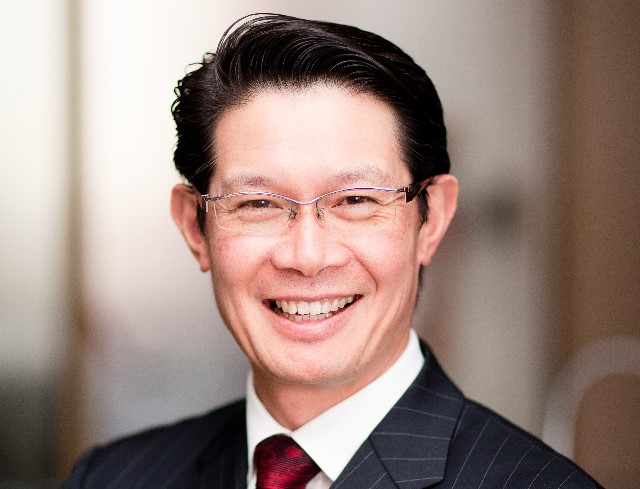
Professor Peter Choong
Head of Department Mentoring and Career Advancement Program
The Department of Surgery runs a focused mentoring and career advancement initiative to support and promote career pathways in academic surgery.The program is directed toward both aspiring and established surgeons and encompasses three main themes.
 Surgery People, Culture and Development
The Department of Surgery is comprised of paid academic surgeons, scientists, and professional staff, honorary staff appointments, with an Executive Committee encompassing representation from all hospital precincts.
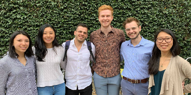 Surgical Students Society
The Surgical Students’ Society of Melbourne (SSSM) is a not-for-profit organisation run by current Doctor of Medicine candidates that represents over 1,400 students across seven clinical school sites in metropolitan Melbourne and rural Victoria.
Still reading?
413
Honours and Masters by Coursework
See our project guide for a wide range of available Honours and Masters projects.
Graduate Research at MDHS
A resource for students and supervisors seeking up to date information about policies and procedures
The University of Melbourne is a globally engaged, comprehensive, research-intensive university uniquely positioned to respond to the major social, economic and environmental challenges of our time.
-
Research Themes
Our research is encompassed within several broad research themes. This multidisciplinary approach aims to increase the opportunities for researchers to collaborate on projects that span common themes.
-
Research Groups
An overview of the research interests within the department and the project work being carried out by our research groups.
Honours and Masters by Coursework
See our project guide for a wide range of available Honours and Masters projects.
Whether you're just starting out or a seasoned professional, we have something to suit your needs and schedule. With the highest teaching evaluations in the Faculty of Medicine, Dentistry and Health Sciences we are proud to offer our coursework and short course programs to our leaders and future leaders.
-
Degrees
The graduate coursework and research degrees offered by Melbourne Medical School are the perfect way to begin or progress your career.
-
Short Courses
Our short courses are designed to keep you up to date with the latest research and information.
-
Scholarships, Bursaries and Prizes
The Faculty of Medicine, Dentistry and Health Sciences offer an extensive range of scholarships and bursaries to undergraduate and postgraduate coursework students.
-
Current Student Resources
Further information for all students in the Melbourne Medical School, including Doctor of Medicine students, plus links to University student resources.

Clinical Electives
View further information for non-University of Melbourne medical students who wish to undertake a clinical elective with one of our hospital partners.
Clinical Schools
The Doctor of Medicine (MD) has been designed to train doctors who have the skills, attributes, passion and competency to make a positive and immediate contribution to health locally and internationally. Integral to your study is the teaching and clinical training you will experience at the University of Melbourne's clinical schools located in affiliated teaching hospitals.
Each of our clinical schools offers our students a unique experience, whilst delivering the same MD curriculum to provide a first-class medical education.
Zone Preferences
Applicants invited to a Multi Mini Interview (MMI) for a place in the Doctor of Medicine will be asked to identify their preference for a particular Clinical School region and whether they are interested in being selected to a particular rural cohort.
The information below should assist applicants in making these decisions.
During the first year of the MD, students are allocated to one of these clinical schools for the final three years of the course (with the exception of non-ERC Rural Clinical School students who may have the opportunity to be re-allocated after completion of Year 2). Acceptance of a place in the medical course indicates acceptance of a place in a particular clinical school zone, and subsequent clinical school placement.
Thank you for engaging with the Faculty of Medicine, Dentistry & Health Sciences. As a friend and supporter of this faculty you will help create scholarships for our students, support groundbreaking research, and build important partnerships and support community engagement.
-
Alumni
Our pride in our graduates and in their lives spent improving the health and well-being of others is matched by our desire to maintain strong connections with all who have passed through our doors.
-
Support
We are grateful for the many ways in which our alumni and donors support the work of the Faculty.
-
Partners
We recognise partnerships and collaborations are an integral facet of our development and a major strength of the school.

Ultrasound Education Group
The Ultrasound Education Group (UEG), University of Melbourne was founded in 2004 by Professors Alistair and Colin Royse. The focus of UEG is on the development of learning in clinical ultrasound as well as hands-on workshops and simulator training programs. UEG also now specialises in the development, publication and delivery of eLearning education courses.
Welcome to the Department of Surgery
Professor Peter Choong, Head of Department
As Head of the Department of Surgery, I am pleased to introduce the Department that forms the focus for academic activities in surgery at the University of Melbourne and the Melbourne Medical School.
Department of Surgery Precincts
The Department is based across seven campuses of the Melbourne Medical School, all at key hospital sites, as well as conducting academic activities in surgery at other hospitals.


Honorary Titles
This section of the Department of Surgery website has been developed to provide a site that collates information that may be of value to our honorary titles – in essence a single point of information.
Diversity and Inclusion
The Melbourne Medical School is proud to foster a vibrant and inclusive culture delivering initiatives that value and support diversity.

DOS Paid Promotions Process and Dates 2023
This section provides further information regarding the Academic Promotions Guidelines and Academic Promotions Timetables for 2023